Publications
SPIE Medical Imaging 2025 Meeting Information
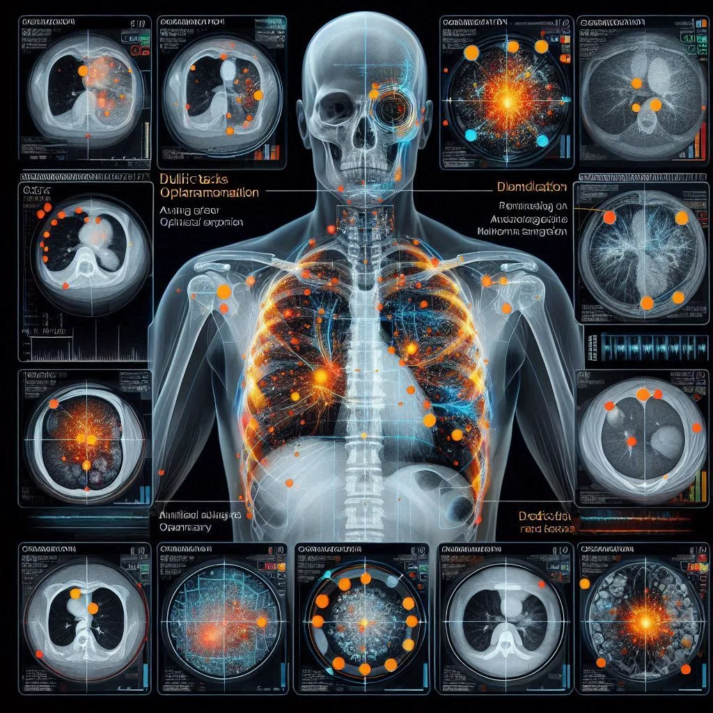
- Automated multi-lesion annotation in chest x-rays: Annotating over 450,000 images from public datasets using the AI-based Smart Imagery Framing and Truthing (SIFT) system
- Development of an AI-based Smart Imagery Framing and Truthing (SIFT) system to annotate pulmonary abnormalities with corresponding boundaries based on CT images

Journal of X-ray Science and Technology
Can AI generate diagnostic reports for radiologist approval on CXR images? A multi-reader and multi-case observer performance study, . 2024 Oct 16. doi: 10.3233/XST 240051.
Abstract :
Background: Accurately detecting a variety of lung abnormalities from heterogenous chest X-ray (CXR) images and writing radiology reports is often difficult and time-consuming.
Objective: To access the utility of a novel artificial intelligence (AI) system (MOM-ClaSeg) in enhancing the accuracy and efficiency of radiologists in detecting heterogenous lung abnormalities through a multi-reader and multi-case (MRMC) observer performance study.

Using artificial intelligence for chest radiograph interpretation: a retrospective multi-reader-multi-case (MRMC) study of the automatic detection of multiple abnormalities and generation of diagnostic report system
In the study, we first introduce a novel AI-based system (MOM-ClaSeg) for multiple abnormality/disease detection and diagnostic report generation on PA/AP CXR images, which was recently developed by applying augmented Mask RCNN deep learning and Decision Fusion Networks. We then evaluate performance of MOM-ClaSeg system in assisting radiologists in image interpretation and diagnostic report generation through a multi-reader-multi-case (MRMC) study.
Using artificial intelligence to assist radiologists in distinguishing COVID-19 from other pulmonary infections
Accurate and rapid diagnosis of coronavirus disease (COVID-19) is crucial for timely quarantine and treatment. Purpose: In this study, a deep learning algorithm-based AI model using ResUNet network was developed to evaluate the performance of radiologists with and without AI assistance in distinguishing COVID-19 infected pneumonia patients from other pulmonary infections on CT scans.
Methods: For model development and validation, a total number of 694 cases with 111,066 CT slides were retrospectively collected as training data and independent test data in the study. Among them, 118 are confirmed COVID-19 infected pneumonia cases and 576 are other pulmonary infection cases.
Impact of artificial intelligence on chest radiography reporting: a multireader-multi-center (MRMC) study of automatic detection of multiple abnormalities and generation of diagnostic report system.
Radiology report writing is a time-consuming task that also requires experience from the radiologists. This study developed and tested a novel AI system (MOM-ClaSeg) to assess its performance in assisting radiologists to automatically detect multiple abnormalities and generate diagnostic report on chest x-ray (CXR).
Using Artificial Intelligence for Chest Radiograph Interpretation: A Prospective Multi-site Study of the Automatic Detection of Multiple Abnormalities and Generation of Diagnostic Report System
Automatic Detection of Multiple Abnormalities and Generation of Diagnostic Report from Radiological Images Using Multi-Task Optimal-Recommendation Max-Predictive Chest X-Ray Classification and Segmentation (MOM ClasSeg) System
Automatic classification of pulmonary abnormalities from chest X-ray images: a multi-task deep learning model
Chest X-ray radiography (CXR) is commonly used to detect different lung abnormalities or diseases. However, interpretation of CXR remains a challenging task for radiologists. In order to overcome this clinical challenge, we develop and test a new MultiTask Optimal-Recommendation Max-Predictive Chest X-Ray Classification and Segmentation (MOM-ClaSeg) system that can detect and delineate 65 different abnormal regions of interest (ROI) on CXR images.

Assessing an AI-based Smart Imagery Framing and Truthing (SIFT) system to assist radiologists annotating lung abnormalities on chest X-ray images for development of deep learning models, Proceedings of SPIE Medical Imaging
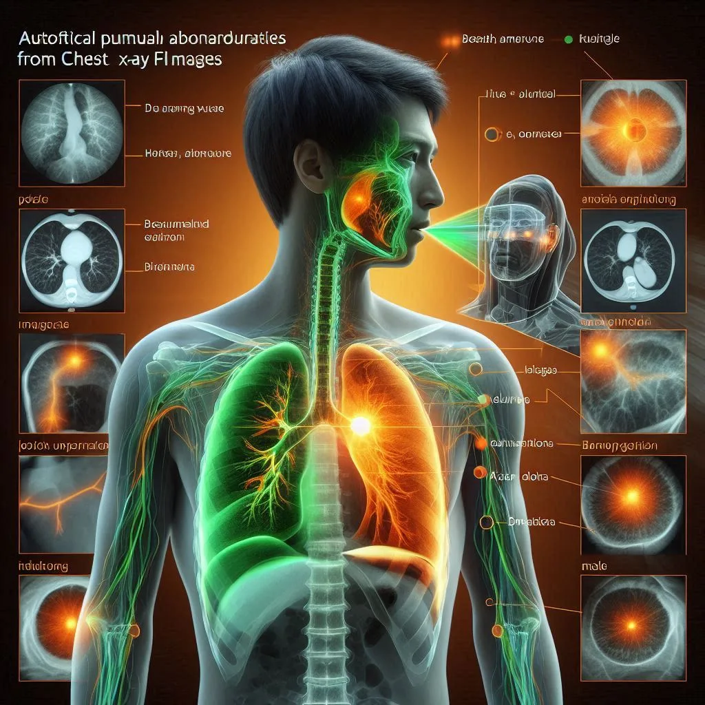
To assess a Smart Imagery Framing and Truthing (SIFT) system in automatically labeling and annotating chest X-ray (CXR) images with multiple diseases as an assist to radiologists on multi-disease CXRs. SIFT system was developed by integrating a convolutional neural network based-augmented MaskR-CNN and a multi-layer perceptron neural network. It is trained with images containing 307,415 ROIs representing 69 different abnormalities and 67,071 normal CXRs. SIFT automatically labels ROIs with a specific type of abnormality, annotates fine-grained boundary.
Developing and assessing an AI-based multi-task prediction system to assist radiologists detecting lung diseases in reading chest x-ray images
Chest x-ray radiography (CXR) is widely used in screening and detecting lung diseases. However, reading CXR images is often difficult resulting in diagnostic errors and inter-reader variability. To address this clinical challenge, a Multi-task, Optimal-recommendation, and Max-predictive Classification and Segmentation (MOM-ClaSeg) system is developed to detect and delineate different abnormal regions of interest (ROIs) on CXR, make multiple recommendations of abnormalities sorted by the generated probability scores, and automatically generate diagnostic report.
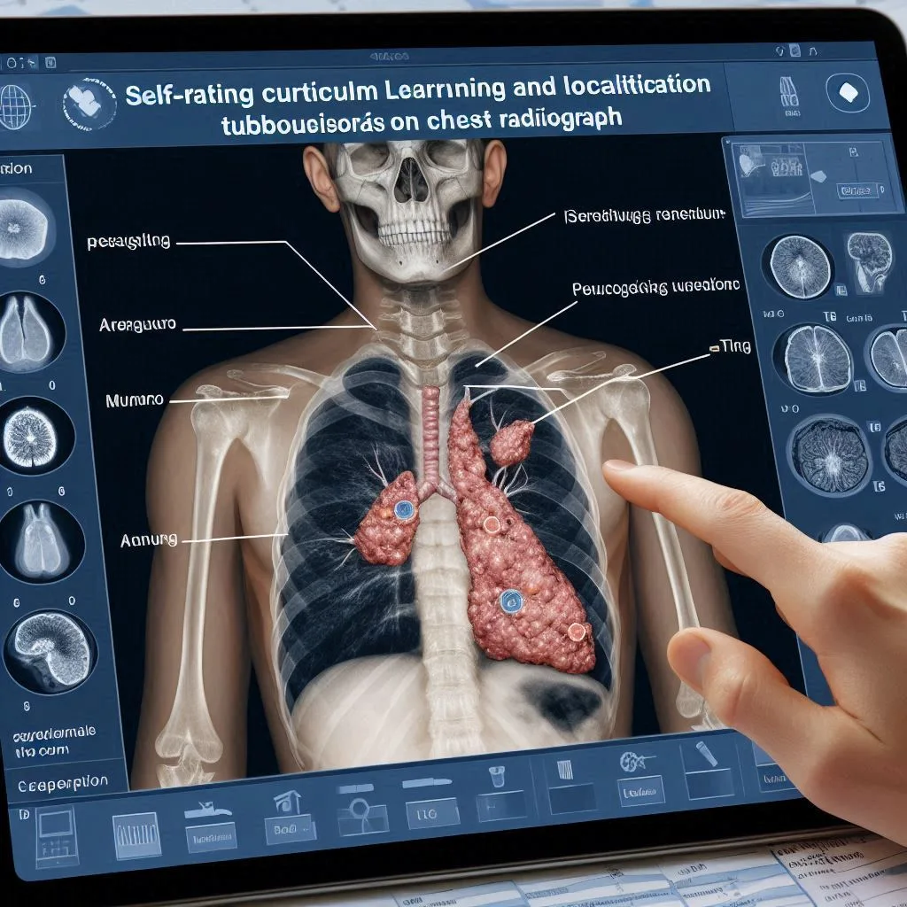
Self-Rating Curriculum Learning for Localization and Segmentation of Tuberculosis on Chest Radiograph
Abstract :
Tuberculosis (TB) is the second leading cause of infectious disease death, and chest X-ray is one of the most commonly used methods to detect TB. In this work, we bring forward a Self-Rating Curriculum Learning (SRCL) method to exploit the task of localization and segmentation of tuberculosis on chest radiographs. A total number of 12,000 CXR images of healthy subjects and bacteriologically-confirmed TB patients, retrospectively collected from multi-center local hospitals, are used in the study.
Get in touch
Accelerate your research and enhance diagnostic accuracy with Caddie's solutions.
Connect with us
ADDRESS:
Ste 300 #1238
Gaithersburg, MD 20879